This article was automatically translated from the original Turkish version.
+1 More
MCF-7
Definition | A breast cancer cell line isolated from a 69-year-old woman in 1970 | ||||||||
|---|---|---|---|---|---|---|---|---|---|
Property | hormone-sensitive ER-positive adenocarcinoma | ||||||||
Field of Use | Gene expression analyses Cancer drug screenings Apoptosis and cell cycle studies | ||||||||
MCF-7 is an estrogen receptor (ER)-positive cell line widely used in human breast cancer research. Developed in 1973 by Dr. Herbert Soule at the Michigan Cancer Foundation, this cell line was isolated from the pleural effusion of a 69-year-old woman of white ethnic origin with metastatic breast cancer. “MCF” is an abbreviation of the institution where the cell line was developed, and “7” indicates it was the seventh cell line generated.
MCF-7 exhibits adherent and semi-suspension characteristics and was derived from a patient with blood group O, Rh+. To date, it has been referenced in more than 25,000 publications and serves as a fundamental model for understanding the biology of ER-positive tumors, hormonal responses, and treatment resistance.
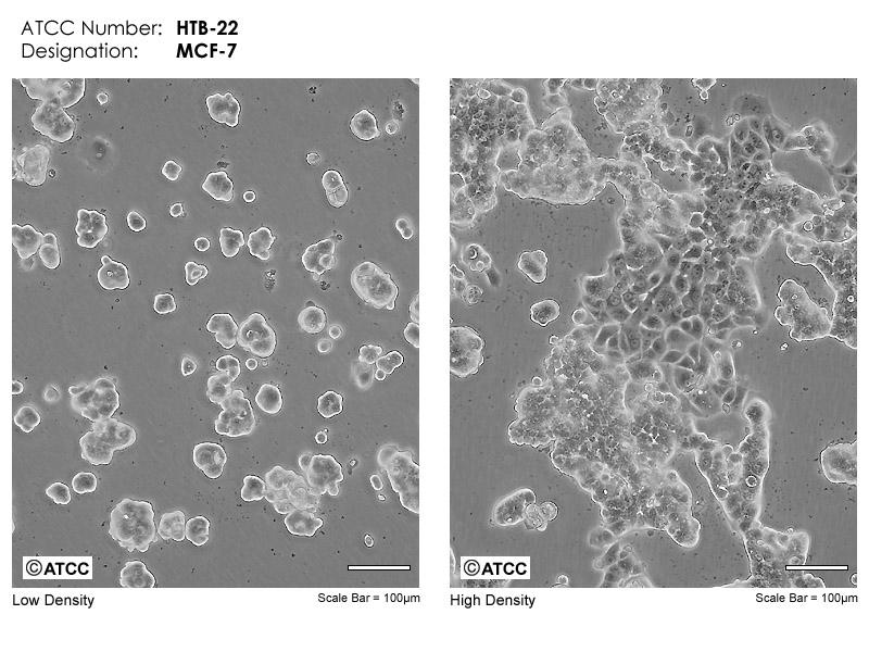
MCF-7 microscopic images(ATCC)
Breast cancer is the most common cancer and one of the leading causes of mortality among women worldwide, treated using various methods including surgery, chemotherapy, radiotherapy, and immunotherapy. However, the development of resistance to chemotherapy, particularly, limits the effectiveness of current treatment approaches and increases the risk of disease recurrence. In this context, photodynamic therapy (PDT) has emerged as a selective and minimally toxic therapeutic approach that targets tumor cells by generating reactive oxygen species (ROS) through the interaction of photosensitizing agents with light and oxygen. Nevertheless, the emergence of therapy-resistant cell populations during PDT applications remains a significant challenge.
In a study conducted at the University of Johannesburg, South Africa, by Aniogo and colleagues, the development of resistance in MCF-7 cells following 10 consecutive cycles of PDT using sulfonated zinc phthalocyanine (ZnPcS4) was investigated. Wild-type (WT) MCF-7 cells were incubated overnight with ZnPcS4 and then exposed to a laser light dose of 20 J/cm². Under these conditions, a 50% cell survival rate was observed in WT cells. Surviving cells were re-cultured and the entire PDT procedure was repeated under identical conditions for a total of 10 cycles. Cells from the 1st, 5th, and 10th generations were separately collected to monitor the resistance development process. The cells obtained after the 10th cycle were designated as “MCF-7/PDT.” The morphological properties, cell cycle (BrdU staining), viability (MTT assay), antioxidant activity (SOD measurement), and P-glycoprotein (P-gp) expression of these resistant cells were evaluated. Results revealed that resistant cells exhibited a mesenchymal phenotype, increased DNA synthesis, and significantly higher P-gp expression compared to parental cells. The data from this study elucidate the biological basis of MCF-7 cell resistance to PDT and provide critical insights for developing more effective future treatment strategies.
In an in vitro study conducted in 2023 at Bolu Abant İzzet Baysal University by Fadime Beyazyüz and colleagues, the antioxidant and anti-proliferative effects of Castanea sativa Mill. (chestnut) leaf ethanol extract were investigated on the MCF-7 human breast cancer cell line. Analyses revealed that the extract had a total phenolic content of 58.22 mg GAE/g, a flavonoid content of 64.62 mg QE/g, and a DPPH radical scavenging activity of 80.06%. The XTT assay for cell proliferation demonstrated time- and dose-dependent cytotoxic effects of the extract on MCF-7 cells, with IC₅₀ values of 100.1 µL at 24 hours, 193 µL at 48 hours, and 15.23 µL at 72 hours. These findings indicate that chestnut leaf extract exhibits significant anti-proliferative and antioxidant effects on MCF-7 cells, likely due to its high phenolic and flavonoid content; however, the authors emphasized that these results require further validation through additional studies.
MCF-7 cells exhibit high expression levels of estrogen receptor alpha (ERα) and progesterone receptor (PR), while ERβ levels are low. HER2 and EGFR receptors are moderately expressed, but HER2 amplification is not observed. These cells belong to the Luminal A molecular subtype. Additionally, these cells express the wnt7b oncogene.
The IGF-1 signaling pathway strongly supports cell proliferation. When applied together with estrogen (E2), IGF-1 exerts a synergistic effect, increasing proliferation by approximately 400–500%. Alone, IGF-1 increases proliferation by about 70%, while E2 increases it by 30%. The cells also express IGFBP-2, IGFBP-4, and IGFBP-5 proteins, whose secretion can be modulated by anti-estrogens.
The modal chromosome number of MCF-7 is 82, ranging from 66 to 87. Karyotype analysis reveals a distribution between hypotriploidy and hypertetraploidy. The 2S component is present at 1%. Each metaphase contains 29–34 marker chromosomes; M1 (submetacentric) and M2–M4 (subtelocentric) chromosomes are observed in over 80% of metaphases. Chromosome 20 is nullisomic, while the X chromosome is disomic.
MCF-7 cells display the following isoenzyme types: AK-1 (type 1), ES-D (type 1–2), G6PD (type B), GLO-I (type 1–2), PGM1 (type 1–2), and PGM3 (type 1). They also retain the ability to metabolize estradiol via cytoplasmic estrogen receptors and to form “dome” structures.
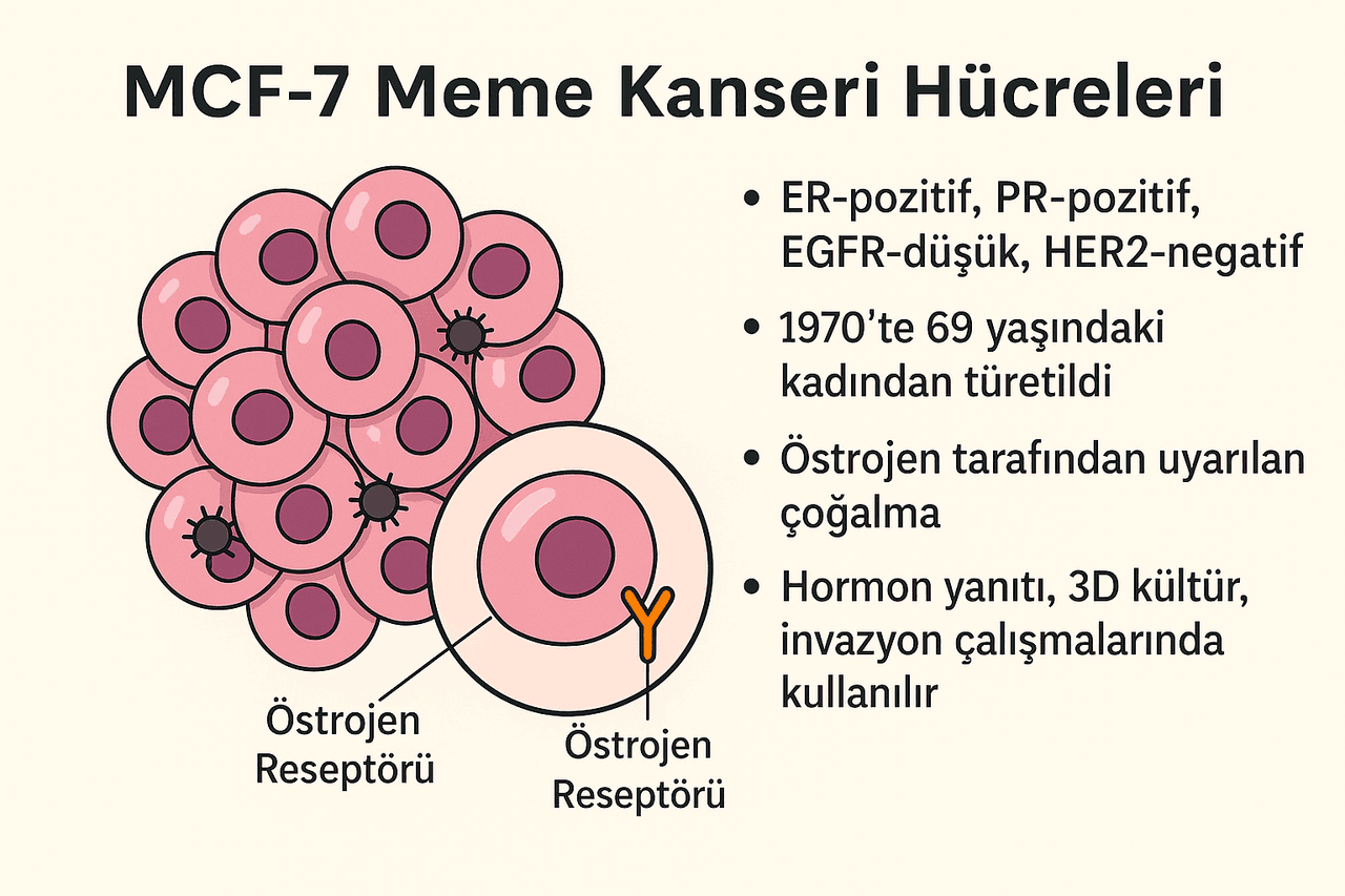
A visual explanation of the MCF-7 cell line (generated by artificial intelligence).
Estradiol promotes cell proliferation by activating proliferative genes such as cyclin D1 via ER. In MCF-7 cells, proliferation increases by approximately 1.3-fold with E2 and 1.7-fold with IGF-1; when both are applied together, proliferation increases 4–5-fold. Anti-estrogen agents (e.g., tamoxifen) inhibit this proliferation by inducing G0/G1 cell cycle arrest; however, long-term exposure may lead to the emergence of resistant subclones. Cell proliferation is also inhibited by TNF-alpha.
Variants developed under long-term estrogen deprivation (LTED) show increased ER expression and become hypersensitive to estrogen. These variants serve as laboratory models for clinical resistance to hormone therapy. In long-term cultures with tamoxifen, the drug’s agonistic effects become dominant, leading to the emergence of “tamoxifen-growing” variants.
Migration and invasion capacity are low in parental MCF-7 cells. However, subclones stimulated by IL-1β (e.g., MCF-7A3) can undergo an epithelial-mesenchymal transition (EMT)-like phenotype characterized by loss of E-cadherin expression and acquire significant invasiveness.
In a study conducted at Biruni University, the chemical composition, antioxidant potential, and anti-proliferative effects of eight different Turkish pomegranate (Punica granatum L.) cultivars were investigated on the MCF-7 human breast cancer cell line. Pomegranate juice extracts were applied at various doses and time points (24, 48, and 72 hours); cell viability was assessed using the WST-1 proliferation assay. Results showed that all extracts reduced MCF-7 cell viability without significant toxicity to MCF-10A normal breast epithelial cells. Notably, the İzmir 1513 (P7) cultivar stood out due to its high anthocyanin (903.66 ± 101.89 μg/g) and punicalagin (992.09 ± 174.53 μg/g) content, exhibiting the strongest cytotoxic effect with an IC₅₀ value of 49.08 µg/ml at 24 hours. These data demonstrate that this study conducted at Biruni University using the MCF-7 cell line supports the potential of Turkish pomegranate varieties as natural anticancer agents.
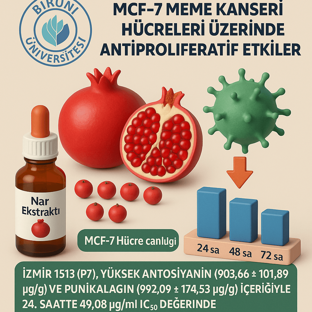
A visual summary of the study (generated by artificial intelligence).
MCF-7 cells contribute minimally to angiogenesis and lymphangiogenesis; levels of VEGF-A, VEGF-C, and VEGF-D secreted by these cells are low. At the receptor level, VEGFR1 and NRP1 are highly expressed, while VEGFR2 is low. However, overexpression of VEGF-C increases lymphatic vessel formation and enhances metastatic capacity, without significantly altering in vitro proliferation.
In 3D culture systems, MCF-7 cells form spheroids. In these spheroids, cells on the outer layers are proliferative, while central cells exhibit hypoxic and apoptotic features. Cell-cell adhesions are mediated by E-cadherin; disruption of these connections abolishes spheroid integrity and organization. Spheroid formation reveals gene expression differences and drug response profiles not observable in 2D culture.
When co-cultured with human mesenchymal stem cells (hMSCs), MCF-7 cells show increased migration and invasion capacity. MCF-7 cells attract MSCs via chemokines, while MSC-secreted growth factors (e.g., TGF-β) disrupt cell organization by suppressing E-cadherin expression. Additionally, MSCs contribute to the formation of capillary-like structures through VEGF.
In a study conducted by researchers from Karadeniz Technical University, the biological effects of Turkish propolis on the human breast cancer cell line MCF-7 were evaluated. The cytotoxic effects of ethanol propolis extract (EEP) were analyzed using the MTT assay; mechanisms such as apoptosis, cell cycle arrest, and mitochondrial membrane potential were examined using flow cytometry, western blot, and spectrophotometric methods. EEP demonstrated selective cytotoxicity against MCF-7 cells, arrested the cell cycle at the G1 phase, increased levels of apoptosis-related proteins p21, Bax, p53, p53-Ser46, and p53-Ser15, and reduced mitochondrial membrane potential. Furthermore, EEP upregulated tumor-suppressive microRNAs such as miR-34, miR-15a, and miR-16-5p, while downregulating the oncogenic miR-21. These findings indicate that Turkish propolis exhibits anti-proliferative and pro-apoptotic effects on MCF-7 breast cancer cells, suggesting its potential as a natural anticancer agent.
The MCF-7 cell line is the gold standard model for studying responses to hormone therapy in ER-positive breast cancers. Resistance to tamoxifen and aromatase inhibitors is linked to epigenetic and transcriptomic alterations and has been extensively characterized in these models. Different subclones of MCF-7 developed in various laboratories (e.g., MCF-7 KO, MCF-7L, MCF-7 ATCC) exhibit distinct receptor levels and drug response profiles, mimicking patient heterogeneity.
Additionally, MCF-7-based cells have been used to understand cross-talk signaling pathways with HER2 and to preclinically evaluate anti-HER2 drugs. Due to these properties, MCF-7 remains an indispensable resource for both basic and translational research.
In a study conducted at the Norwegian Radium Hospital, the binding, internalization, and cytotoxic effects of alpha particle-emitting 227Th-trastuzumab were evaluated in breast and ovarian cancer cell lines overexpressing HER2. In this study, the MCF-7 cell line, which exhibits low HER2 expression, was used as a control group, with comparative analyses performed against BT-474, SKBR-3, and SKOV-3 cell lines. Approximately 15-fold more HER2 antigen regions were detected in BT-474 and SKBR-3 cells compared to MCF-7; fluorescence microscopy confirmed internalization and vesicular localization of trastuzumab. High levels of surface and internalized radioactivity were observed in BT-474 and SKBR-3 cells, whereas uptake in MCF-7 cells remained limited. After 1 hour of 227Th-trastuzumab application, the average absorbed dose was calculated at 2–2.5 Gy, which dose-dependently suppressed cell growth and induced apoptosis. The findings demonstrate that 227Th-trastuzumab produces stronger and more selective cytotoxic effects in HER2-positive cells than X-ray irradiation.
In a similar study conducted at Yeditepe University in Türkiye, microRNAs potentially involved in regulating the response to trastuzumab therapy in HER2-positive breast cancer cells were investigated. The MCF-7 cell line was used as a control group, with comparative analyses performed against BT-474 and SK-BR-3 cell lines. qRT-PCR array analysis after tamoxifen and trastuzumab treatments revealed a significant increase in miR-770-5p expression, independent of cell type or drug. Overexpression of this miRNA reduced cell motility and invasion capacity in HER2+ cells, with a marked decrease in HER2 protein levels observed particularly in BT-474 cells. Furthermore, expression of both total and phosphorylated forms of AKT and ERK, key components of the PI3K and MAPK signaling pathways, was reduced, indicating that miR-770-5p enhances the anti-proliferative effect of trastuzumab. The study suggests that modulation of miR-770-5p expression could represent a potential strategy to improve the efficacy of trastuzumab therapy.
ATCC. “HTB-22 (MCF-7) [Cell Line].” American Type Culture Collection. Accessed July 23, 2025. https://www.atcc.org/products/htb-22
Adrian V. Lee, Steffi Oesterreich, Nancy E. Davidson
Aniogo, Eric Chekwube, Blassan P. George, and Heidi Abrahamse. "Characterization of Resistant MCF-7 Breast Cancer Cells Developed by Repeated Cycles of Photodynamic Therapy." Frontiers in Pharmacology 13 (September 16, 2022). https://doi.org/10.3389/fphar.2022.964141
Beyazyüz, F., E. Gülbahçe Mutlu, S. Alpa, F. Z. Erbayram, F. N. Türkoğlu, and Ş. Kulaç. "Determination of Antioxidant and Anticancer Activities of C. Sativa Leaf Extracts on MCF7 Human Breast Cancer Cell Line." Medical Records 5, no. 3 (September 2023): 472–477.
CancerTools.org. "MCF7/ExeR-3 Cell Line – Cat. #152557." Cancer Research UK Reagents and Tools Portal. Accessed July 23, 2025. https://cancertools.org/cell-lines/mcf7-exer-3-152557/
Comșa, Șerban, Anca Maria Cîmpean, and Marius Raica. 2015. "The Story of MCF-7 Breast Cancer Cell Line: 40 Years of Experience in Research." Anticancer Research 35 (6): 3147–3154. Date Published: June 2015.
Date Published: March 31, 2015
Eroglu Ozkan, Esra, Mehmet Fatih Seyhan, Ozlem Kurt Sirin, Tugba Yilmaz-Ozden, Ezgi Ersoy, Seda Damla Hatipoglu Cakmar, Ahmet Ceyhan Goren, Hulya Yilmaz Aydogan, and Oguz Ozturk. “Antiproliferative Effects of Turkish Pomegranate (Punica granatum L.) Extracts on MCF-7 Human Breast Cancer Cell Lines with Focus on Antioxidant Potential and Bioactive Compounds Analyzed by LC-MS/MS.” Journal of Food Biochemistry 45, no. 9 (August 19, 2021): e13904. https://doi.org/10.1111/jfbc.13904
Heyerdahl, H., C. Krogh, J. Borrebæk, Å. Larsen, and J. Dahle. "Treatment of HER2-Expressing Breast Cancer and Ovarian Cancer Cells with Alpha Particle-Emitting 227Th-Trastuzumab." International Journal of Radiation Oncology, Biology, Physics 79, no. 2 (February 1, 2011): 563–570.
JNCI: Journal of the National Cancer Institute, Volume 107, Issue 7, July 2015, djv073, https://doi.org/10.1093/jnci/djv073
MCF-7 Cells—Changing the Course of Breast Cancer Research and Care for 45 Years
Misir, S., Y. Aliyazicioğlu, S. Demir, İ. Turan, and C. Hepokur. "Effect of Turkish Propolis on miRNA Expression, Cell Cycle, and Apoptosis in Human Breast Cancer (MCF-7) Cells." *Nutrition and Cancer* 72, no. 1 (2019): 133–145. https://doi.org/10.1080/01635581.2019.1616100
Noyan, Senem, Hakan Gurdal, and Bala Gur Dedeoglu. "Involvement of miR-770-5p in Trastuzumab Response in HER2 Positive Breast Cancer Cells." PLOS ONE, April 22, 2019. https://doi.org/10.1371/journal.pone.0215894
MCF-7
Definition | A breast cancer cell line isolated from a 69-year-old woman in 1970 | ||||||||
|---|---|---|---|---|---|---|---|---|---|
Property | hormone-sensitive ER-positive adenocarcinoma | ||||||||
Field of Use | Gene expression analyses Cancer drug screenings Apoptosis and cell cycle studies | ||||||||
Molecular and Cellular Characteristics
Proliferation, Migration, and Treatment Response
Microenvironment, 3D Architecture, and hMSC Interactions
Clinical Significance and Applications